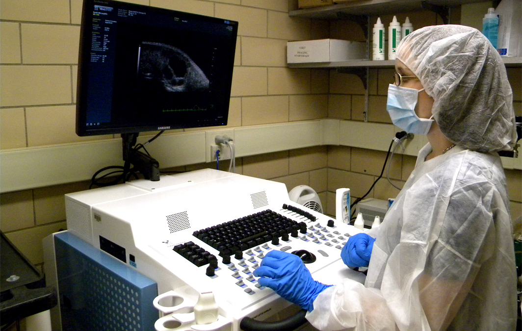
The Animal Models & Imaging Facility (AMIF) provides resources for small animal imaging and technical support for preclinical research models. In vivo imaging modalities include 2D optical imaging or 3D optical tomography for measuring bioluminescence and fluorescence signals, high frequency micro-ultrasound imaging and small animal MRI imaging. A high resolution microCT is available for ex vivo
samples. The facility also includes systems for body composition or metabolic analyses, as well as a small animal X-ray irradiator.
Equipment
- PerkinElmer IVIS SpectrumCT
- Bruker SkyScan 1272 MicroCT
- VisualSonics Vevo F2
- Xstrahl XenX
- Columbus Instruments CLAMS
- EchoMRI
- Aspect M7 MRI
- Workstation #3
Contacts
Director
Karen Martin, PhD | kamartin@hsc.wvu.edu
Manager
Amanda Stewart, PhD | abarker@hsc.wvu.edu
Imaging Specialist
Sarah McLaughlin | smclaughlin@hsc.wvu.edu
Recent News
Read the 2023 Imaging Facilities Newsletter.
Acknowledging the Animal Models & Imaging Facility
Please remember to acknowledge support for AMIF in all your publications:
Imaging experiments and image analysis were performed in the West Virginia University Animal Models & Imaging Facility which has been supported by the WVU Cancer Institute, the WVU HSC Office of Research and Graduate Education, and NIH grants P20GM121322 and U54GM104942.
If you make use of the following equipment, please include these funding sources in your acknowledgement:
- IVIS SpectrumCT: U54GM104942
- VisualSonics Vevo 2100: S10RR026378
- VisualSonics Vevo F2: P20GM144230
- Xstrahl XenX Irradiator: U54GM104942
- Workstation #3: P30GM103488
